Figure 2.
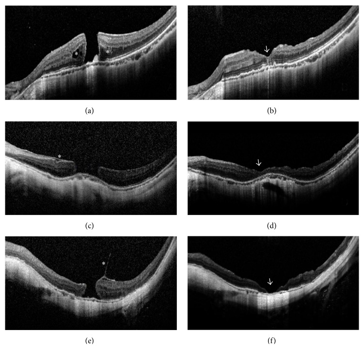
Optical coherence tomography (OCT) images of large myopic macular holes (MHs) in selected cases before and after surgery. (a) Case Number 5 before surgery. An MH of 563 μm and retinoschisis around the MH (asterisks) was present. Best-corrected visual acuities (BCVAs) = 0.02. (b) Case Number 5 at 6 months after surgery. Type 1 closure in U-shape and restoration of the inner and outer segment junction line was achieved (arrowhead). BCVA = 0.3. (c) Case Number 12 before surgery. A large-sized MH of 812 μm and epiretinal membrane (asterisk) was present. BCVA = FC/50 cm. (FC = finger counting) (d) Case Number 12 at 6 months after surgery. Type 1 closure in shallow V-shape was present (arrowhead). BCVA = 0.3. (e) Case Number 14 before surgery. A longstanding MH of 491 μm and vitreomacular traction (asterisk) was present. BCVA = HM/BE. (HM = hand motion; BE = before eye) (f) Case Number 14 at 6 months after surgery. Type 2 closure with bare retinal pigment epithelium in the center (arrowhead) was present. BCVA = HM/BE.

