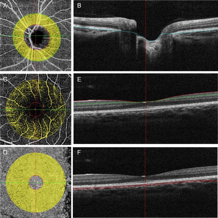
Figure 1.
Peripapillary and parafoveal perfusion was measured by optical coherence tomography angiograms of the emmetropic eye. (A) The peripapillary region was defined as a 700 µm wide elliptical annulus extending outward from the optic disc boundary in optical coherence tomography (OCT) retinal angiograms. (B) The boundaries used for segmentation are indicated by the blue lines (retinal pigment epithelium) on cross-sectional OCT reflectance. (C and D) A masking procedure of measuring parafoveal retinal (C) and choroidal (D) perfusion consisted of an annulus defined by an inner diameter of 0.6 mm and an outer diameter of 2.5 mm. (E) The superficial area of parafovea was defined as from inner limiting membrane with offset of 3 µ to inner plexiform layer with offset of 29 µ. (F) The choroid area of parafoveal was defined as from retinal pigmental epithelium reference with offset of 29–59 µ.

