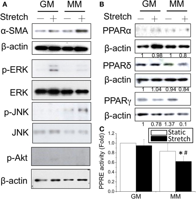
Figure 3.

Application of cyclic stretch (10% strain at 1 Hz for 1 h) in MM with further induced myogenesis as indicated by increased α-SMA expression (A). The phosphorylation of JNK was induced by applying stretch in MM, whereas ERK phosphorylation was observed when stretch was applied in GM. A decrease in PPARγ protein expression was also found during stretch-induced myogenesis in MM (B). The number below each lane indicates the quantified fold change with normalized to GM static condition and its individual β-actin. A further decrease of PPRE promoter activity was detected when applying cyclic stretch (C). *Significant difference compared with GM (p < 0.05). #Significant difference compared with MM under static conditions (p < 0.05).
