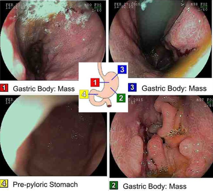
Figure 3.
Esophagogastroduodenoscopy with marked images of where the picture was taken along the tract. Findings are significant for a normal oesophagus. Also note the malignant gastric tumour in the cardia, in the gastric fundus, in the gastric body, on the anterior wall of the stomach, on the greater curvature of the stomach, on the lesser curvature of the stomach, and on the posterior wall of the stomach and at the incisura. The duodenum was normal.

