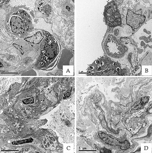
Figure 3.

Ultrastructural preservation of FSE Epon embedded tissues. A-B) Lung tissues processed by standard transmission electron microscope protocol (5000x). C-D) Lung samples processed by FSE method showed a satisfactory preservation of ultrastructural details. Indeed, for lower magnifications (<10,000x), it was still possible to carry out morphological analysis (5000x).
