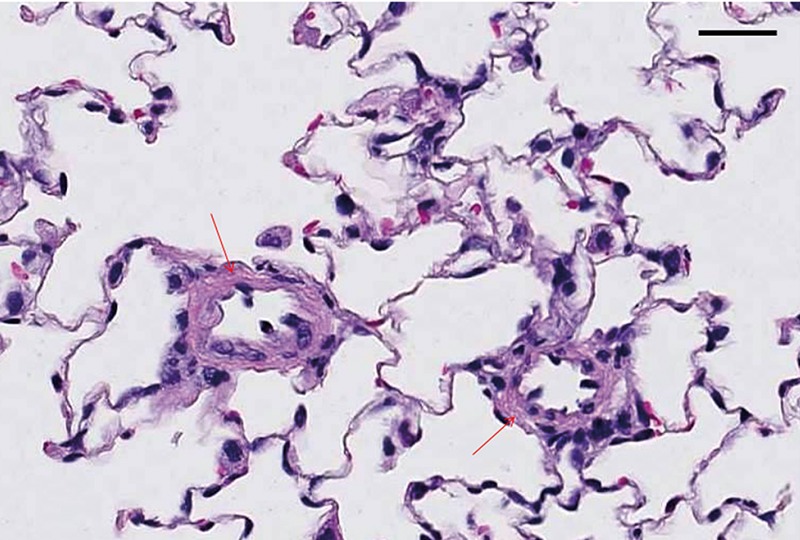
Figure 1.

Rat lung tissue section was fixed in 10% NBF for 48 h followed by 70% EtOH for 5 days and stained with hematoxylin and eosin, sectioned at 4 µm. Note small caliber vessels in MCT-induced PAH lung showing the pathology of increased muscularization of small caliber vessel walls (arrows). Scale bars: 25 µm.
