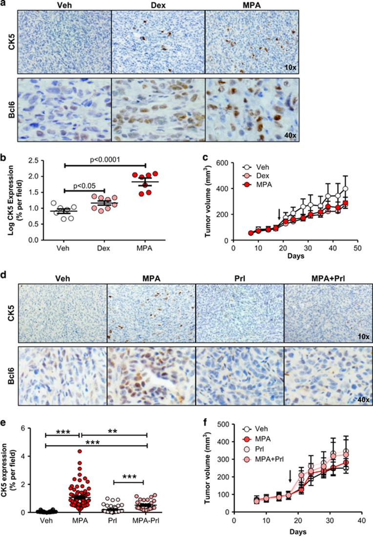
Figure 6.
Dexamethasone and medroxyprogesterone acetate treatment induces CK5 and BCL6 expression in vivo, and is suppressed by prolactin. (a) Representative images of T47D xenograft tumors harvested from mice implanted with Veh, Dex or MPA slow-release pellets for 28 days and subjected to immunohistochemistry for CK5 (top) and BCL6 (bottom) using DAB chromogen. (b) Scatter dot plots of the log percent area of CK5+ cells within these tumors. (c) Tumor growth plots in tumor-bearing mice treated with Veh, Dex or MPA as indicated. (d) Representative images of T47D xenograft tumors harvested from mice implanted with slow-release pellets of MPA or Veh with and without co-treatment with biweekly injections of prolactin (Prl) for 21 days and subjected to immunohistochemistry for CK5 (top) and BCL6 (bottom). (e) Scatter dot plots of percent area of CK5+ cells in images taken of these tumors (mean±s.e.m. indicated). (f) Tumor growth plots in mice treated with Veh, MPA, Prl or MPA+Prl as indicated. Arrow indicates initiation of treatment on day 18. *P<0.05; **P<0.01; ***P<0.001.

