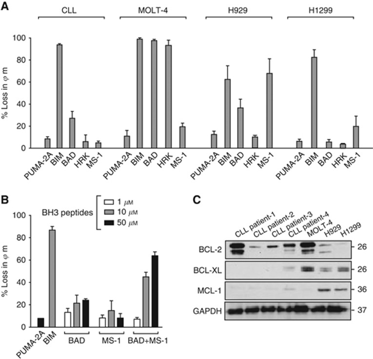
Figure 1.
BH3 profiling in cell lines. (A) Cells were incubated with BH3 peptides (10 μM) in TEB buffer (containing 0.002% digitonin) for CLL cells (30 min), MOLT-4 cells (1 h), and H929 and H1299 cells (2 h). Mitochondrial potential was assessed and changes calculated with reference to DMSO & FCCP-treated cells. Data represents the Mean±s.e.m. of triplicate experiments. (B) Mitochondrial depolarisation of CLL cells exposed to BAD and MS-1 peptides alone or in combination. (C) Western blots of either CLL cells from four patients or the cell lines were analysed for expression of the indicated proteins. No detectable BCL-w or BFL-1 was observed in any of the cells.

