. 2015 Dec 23;7(3):2392–2398. doi: 10.1039/c5sc03717d

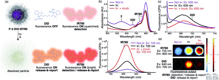
Fig. 4. Increase in fluorescence of DiD and IR780 upon light-triggered release from P-2 particles indicates particle status. (a) Scheme describing the fluorescent state of DiD and IR780 encapsulated in P-2-DiD-IR780 particles before (top) and after irradiation with red light (bottom). (b) Absorbance of P-2-DiD-IR780 particles (2: 9 × 10⁻⁵ M, 1 mL, DiD: 1 × 10⁻⁶ M (1 mol% loading compared to 2), IR780: 5 × 10⁻⁶ M (6 mol% loading compared to 2), in H2O with 1% w/v poloxamer 407) before (purple trace), after 7.5 min irradiation (λex > 590 nm, 183 mW, 65 mW cm–2, black trace) and after irradiation and 10 min incubation at 37 °C (red trace). (c–d) Fluorescence of the same samples excited at (c) 635 nm (DiD channel) and (d) 720 nm (IR780 channel). (e) IVIS image of a well plate containing a similar P-2-DiD-IR780 dispersion before irradiation (left well), after irradiation (middle well) and after irradiation and incubation (right well). Top, IR780 fluorescence channel (λem = 800 nm); bottom, DiD fluorescence channel (λem = 700 nm). Both rows are the same three wells.

Fig. 4