Figure 1.

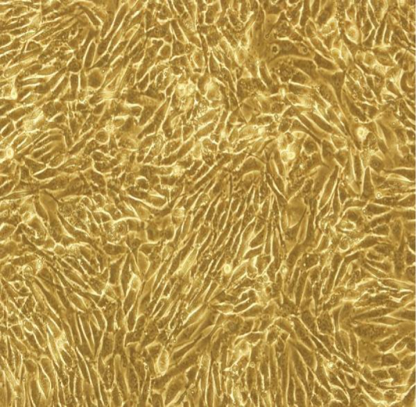
Phase contrast microscopy of hCMEC/D3 (200 × ). The cells were cultured in endothelial growth medium-2 and attained confluence after 3-5 days.

Phase contrast microscopy of hCMEC/D3 (200 × ). The cells were cultured in endothelial growth medium-2 and attained confluence after 3-5 days.