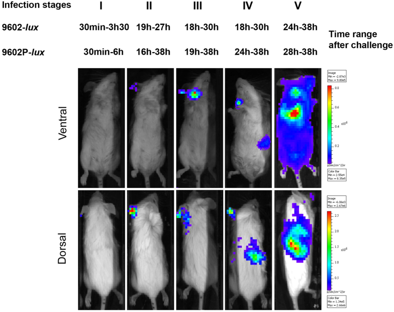
Figure 1. Cutaneous anthrax is stratified along five stages through the time pattern of bioluminescent signalling.
BALB/c mice were infected into the ear pinna with spores of the bioluminescent B. anthracis strains - 2.73 ± 0.32 log10 wild type 9602-lux (mean ± SD, n = 24) or 2.46 ± 0.35 log10 pagA-deleted 9602P-lux (mean ± SD, n = 19), as described in the Material and Methods section - and the dynamics of infection followed for each mouse through BLI. Images in this figure depict photographs overlaid with false-colour representations of luminescence intensity, where red is most intense and blue is least intense. Top row: ventral view of a single representative mouse at the indicated time range after infection. Bottom row: dorsal view of the same mouse. Five stages were distinguished (noted I to V) with the time range for all the mice belonging to each stage. The earliest time of infection was associated with absence of BLI (stage I). Stages II, III and IV corresponded to BLI signal displayed in specific organs targeted by B. anthracis, i.e. the inoculated ear (stage II), the efferent draining cervical lymph nodes (stage III) and the spleen (stage IV). The final stage of infection associated with lung BLI and septiceamia was noted stage V. The image series are representative of at least 5 independent animal groups for each time range after challenge.

