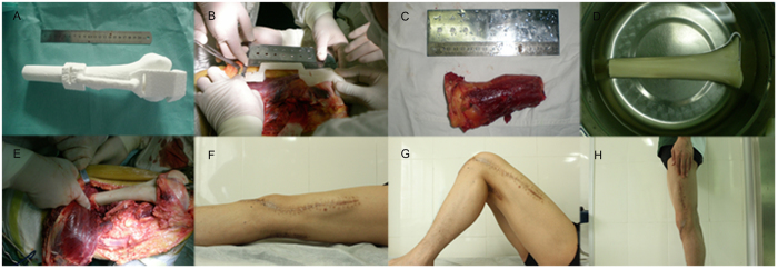
Figure 6. The guiding template applied in the operation on an 18-year-old man with a distal femur.
(A) 3D printed guiding template fitted with the femur model. (B) The guiding template fitted perfectly with the distal femur during the operation and provided anatomical guidance for a safe osteotomy line. (C) Precise excision of the tumorous bone according to the surgery guided by the template. (D) The pruned allografted bone to be implanted. (E) The allograft was implanted into the bone defect. (F–H) The patient was healed (F, Supine view; G, Flexion view; H, Standing view).

