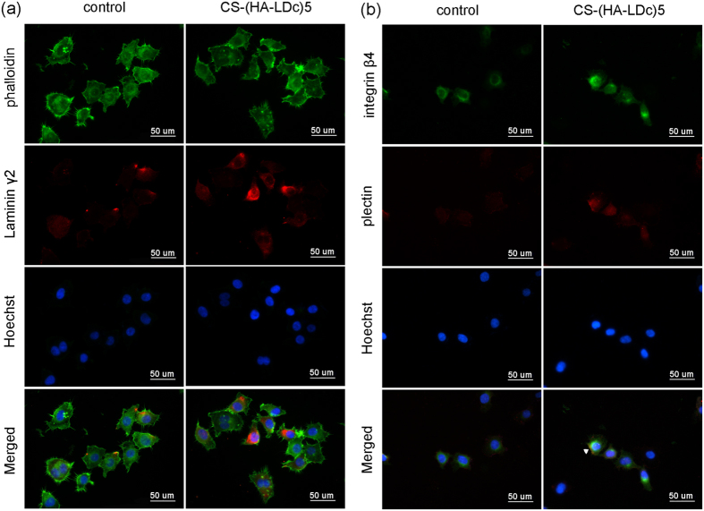
Figure 5. Effects of CS-(HA-LDc)5 coating on the expression of laminin γ2 and hemidesmosomal components in HN4 cells.
(a) Immunofluorescent microscopy expression of laminin γ2 in HN4 cells (red); cytoskeleton (green); nuclei (blue). (b) Integrin β4 was in green, plectin was in red and nuclei were in blue. Co-localized staining in the merged image appeared in yellow, indicating the HD-like structure (white arrow head). Samples were examined under ×400 magnification.

