Figure 1.
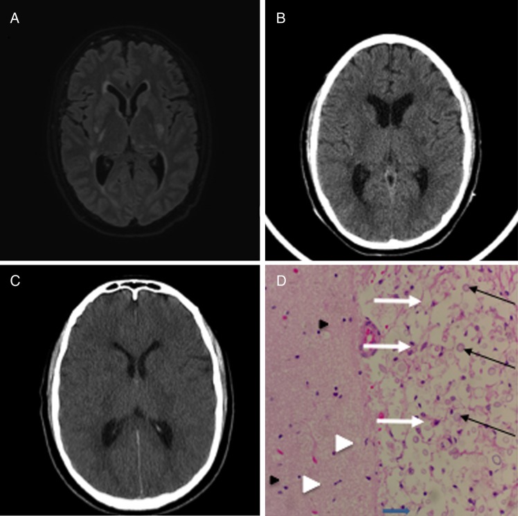
(A) Axial noncontract 3D FLAIR-SENSE magnetic resonance image, hospital day 1, showing areas of hyperintensity in the basal ganglia bilaterally representing ischemic infarcts due to cryptococcal vasculopathy. (B) Axial noncontrast computed tomography (CT) head, hospital day 9, after the initial seizure-like event. (C) Axial noncontrast CT head, hospital day 10, after the acute decline in neurologic status showing the interval development of diffuse cerebral edema. (D) Representative hematoxylin and eosin stain of cerebral white matter from the frontal lobe showing Cryptococcus (black arrows) with classic “soap bubble” appearance and surrounding tissue destruction. Microglia (white arrowhead) and reactive astrocytes (white arrows) are also present. The left side shows intact neuropil (pink, strands in the background) as well as more microglia and scattered oligodendrocytes (black arrowhead).

