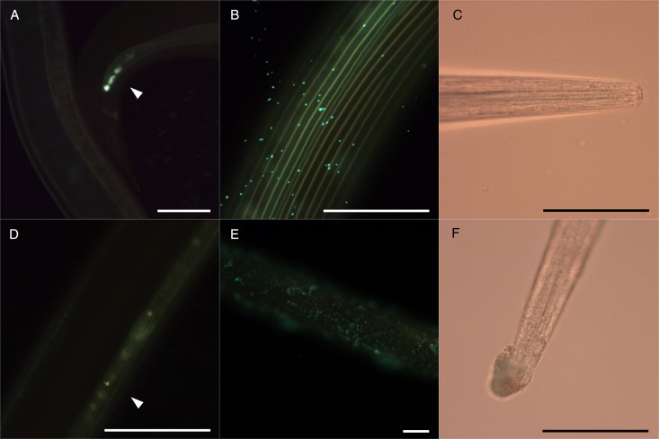
Fig. 6.
Micrographs of Ostertagia ostertagi adults pre-incubated in control media (A, B, C) or CT fraction from blackcurrant leaves A at 300 µg mL−1 (D, E, F), for 2 h (A, D) or 30 h (B, C, E, F), and then transferred to control media containing fluorescent E. coli for 24 h. White arrow heads show presence of fluorescent bacteria in the cloacae (A) or in the digestive tract (B) of the worm. (B, E) show a part of the cuticle and (C, F) the anterior part (scale bars = 100 µm).

