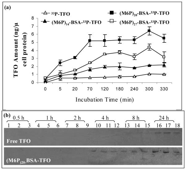
FIGURE 1.
Cellular uptake of M6P-BSA-TFO by HSC-T6 cells. (A) Time profile of the cellular uptake of M6P-BSA-33P-TFO and 33P-TFO by HSC-T6 cells. HSC-T6 cells were incubated with 33P-TFO or M6P-BSA-33P-TFO (3×10⁴ cpm, 0.4 μM) at 37 ºC and then cells were harvested at indicated time for measurement of associated radioactivity. TFO concentration was expressed in terms of ng TFO/μg cell protein. Values are mean ± SD of 3 independent experiments. (B) Polyacrylamide gel electrophoresis (PAGE) of the TFO extracted from HSC-T6 cells. HSC-T6 cells were incubated with 33P-TFO or (M6P)20-BSA-33P-TFO at 37 ºC for up to 24 h. Cells were harvested at indicated time and lysed with 1% SDS lysis buffer. TFO was extracted and analyzed by gel electrophoresis using a 20% polyacrylamide gel. Each time point was presented with 3 independent experiments.

