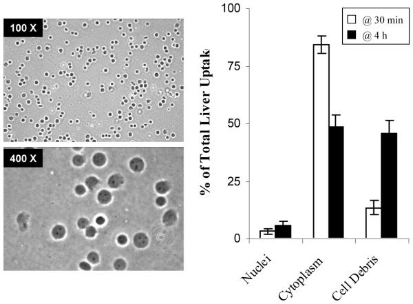
FIGURE 8.
Subcellular distribution of (M6P)20-BSA-33P-TFO in the liver. At 30 min and 4 h after intravenous administration of (M6P)20-BSA-33P-TFO into normal rats at a dose of 0.2mg/kg, rats were sacrificed and the livers were removed. The nuclei, cytoplasm and cell debris were isolated from liver tissues using a sucrose gradient. Distribution in the nuclei, cytoplasm and cell debris were given as % of the total liver uptake and values are mean ± SD of 4 rats in each group.

