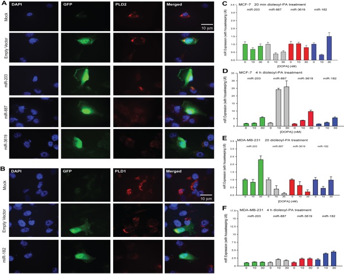
FIG 6.
PLD2 protein expression decreases in the presence of miRNA in MDA-MB-231 breast cancer cells, and miRNA gene expression increases in response to PA. (A, B) Immunofluorescence of cells transfected with plasmids containing an miRNA gene and a GFP gene. miRNA gene expression was represented by GFP expression. The empty plasmid backbone contained the GFP gene and no miRNA gene. PLD2 and their targeting miR-203, miR-887, and miR-3619-5p (A) or PLD1 and its targeting miR-182 (B) were represented with TRITC. (C to F) Gene expression of miR-203, miR-887, miR-3619-5p, and miR-182 after incubation with increasing amounts of DOPA (1,2-dioleoyl-sn-glycero-3-phosphate) for 20 min or 4 h in MCF-7 or MDA-MB-231 cells. Note that the two y axis scales in panels D and F are different since expression in MCF-7 is lower than in MDA-MB-231 cells.

