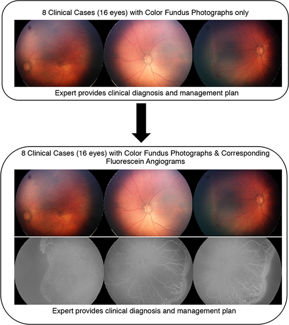
Figure 1. Study Design Presented to Retinopathy of Prematurity (ROP) Experts to Determine ROP Diagnosis and Management Plan using Color Fundus Photography and Fluorescein Angiography (FA).
In the first part of this study, each expert completed 8 clinical cases in which he or she provided a diagnosis of zone, stage, plus, category, management, advanced posterior-ROP, and recommended clinical follow-up. In the second part of the study, experts were presented with the same 8 clinical cases, but were now provided with the corresponding FAs. For each clinical case, the experts were asked to provide a clinical diagnosis and management plan, but were not able to see their previous responses from the first part of the study.

