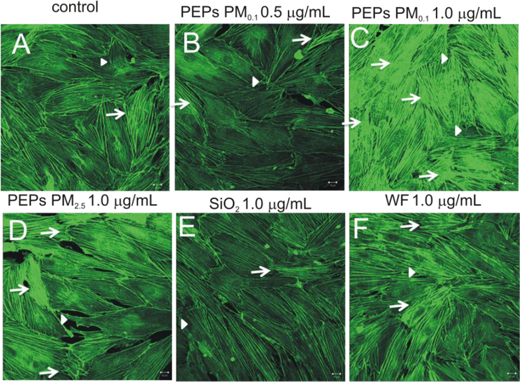
Figure 3. SAEC exposure to PEPs increases actin filament remodeling in HMVEC.
HMVEC were grown on coverslips in the co-culture model and stained with AlexaFluor 546. (A) Control, (B) PEPs PM0.1 0.5 µg/mL, (C) PEPs PM0.1 1.0 µg/mL, (D) PEPs PM2.5 1.0 µg/mL, (E) SiO2 1.0 µg/mL, and (F) WF 1.0 µg/mL. Arrows represent increase actin-filament stress fibers and arrowheads indicate increase filopodia and lamellipodia. Images were taken using a Zeiss LSM510 microscope. Images are a presentation of n = 3.

