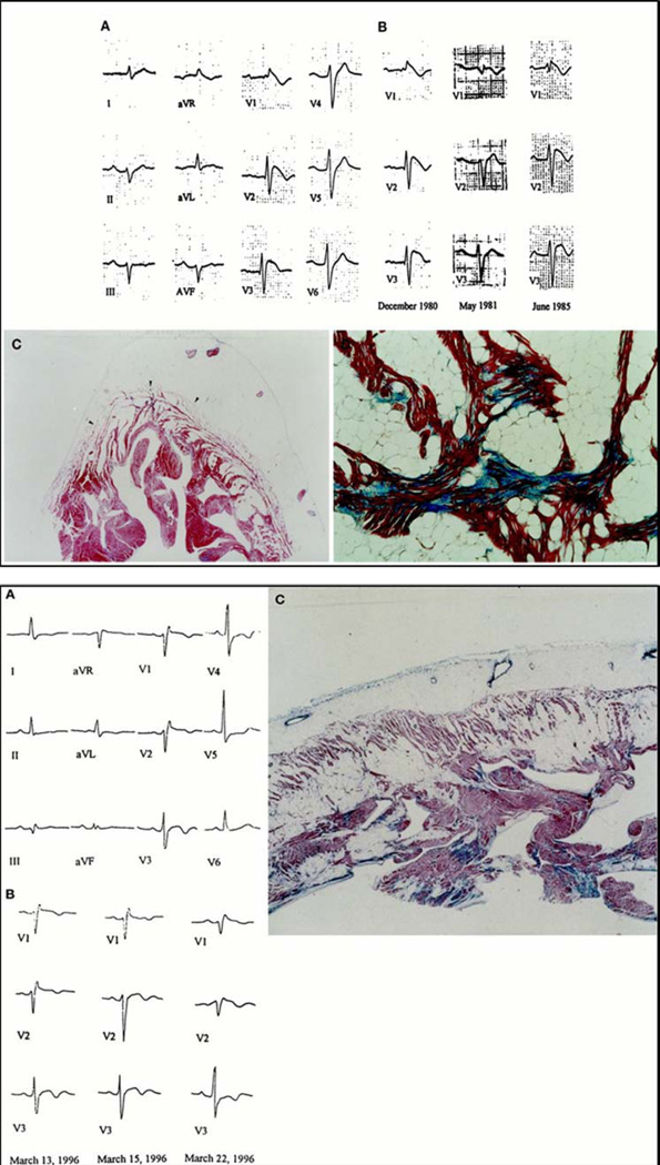
Figure 3.
Electrocardiographic and histopathologic findings in two sudden death victims with overlapping phenotype of ARVC and BS. Top: A 35 year-old man who died suddenly at rest. Baseline 12-lead ECG showing first degree AV block and a coved-type Brugada ECG (A). Note the serial changes over years of right precordial repolarization abnormalities with transient near-normalization (B). Panoramic histological view of RV free wall showing transmural myocardial loss with fatty replacement, mostly involving subepicardial and midmural layers (C). At higher magnification, histological examination of RV myocardium shows tiny interstitial fibrosis in the setting of myocardial atrophy and fatty tissue (D). Bottom: A 27 year-old who died suddenly while sleeping. Baseline 12-lead ECG showing a coved-type Brugada ECG (A). Day-to-day changes of right precordial ST-segment pattern, which exhibits maximum displacement upward on March 22, 1996 (B). Panoramic histological view of RV myocardium disclosing full-thickness fibrofatty myocardial replacement (C).
Adapted from Corrado et al.45

