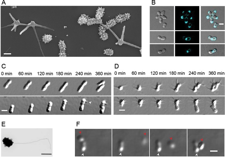
FIG 1.
P. hirschii has two distinct morphotypes. (A) Scanning electron microscope image of P. hirschii cells highlights the short- and long-stalked morphologies. The image was acquired at 60,000× magnification. Scale bar = 1 μm. (B) Fluorescent d-amino acid staining of cells reveals polar and midcell peptidoglycan synthesis. Scale bar = 2 μm. (C) Time-lapse differential interference contrast (DIC) images taken every 60 min on MMB agar pads show a short-stalked mother cell giving rise to a short-stalked daughter cell (top) and a short-stalked mother cell giving rise to a long-stalked daughter cell (bottom). The white arrowhead indicates the formation of a long stalk. Scale bar = 2 μm. (D) A long-stalked mother cell gives rise to a long-stalked daughter cell (top), and a long-stalked mother cell gives rise to a short-stalked daughter cell (bottom). Scale bar = 2 μm. (E) Transmission electron micrograph of an individual short-stalked P. hirschii cell with a single polar flagellum. Scale bar = 1 μm. (F) Montage showing a nonmotile mother cell producing a motile daughter cell. The white arrowheads indicate the stationary mother cell. The red arrowheads indicate the position of the motile daughter cell in the each image. The images shown were acquired at 20-min intervals. Scale bar = 2 μm.

