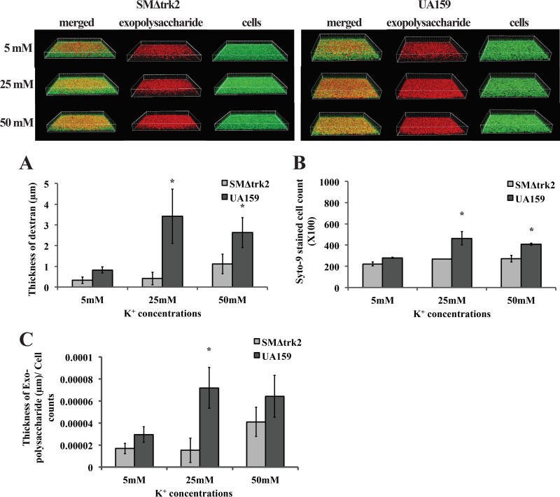
FIG 6.
Confocal laser scanning microscopy of biofilms formed by S. mutans wild-type strain UA159 and trk2-null mutant strains. Biofilms were grown on an 8-well chambered cover glass containing MMSK supplemented with 5 mM, 25 mM, or 50 mM KCl and 1 μM Texas Red-labeled dextran conjugate. At 18 h, all of the bacterial cells in the biofilms were labeled with Syto 9 green fluorescent nucleic acid stain and processed for confocal laser scanning microscopy (CLSM). The histograms represent the average surface thickness of dextran (A), average spot counts of objects in the biofilms (B), and the dextran thickness normalized to the Syto 9-stained-cell count (C). Values are means and standard errors (SE) from experiments repeated three times with three technical replicates, and statistical comparison was performed using Student's t test. A P value of <0.05 was considered significant (*).

