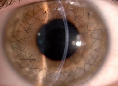
Figure 3.

Biomicroscopic examination showed a clear and well integrated lamellar graft in a 35-year-old keratoconus patient who had descemetic deep anterior lamellar keratoplasty (big-bubble technique) 6 months previously. Two double-running 12 bites sutures are present.
