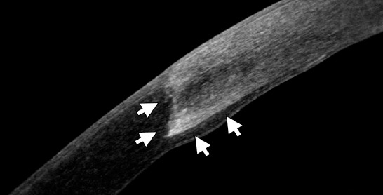
Figure 4.

Anterior segment Fourier-domain optical coherence tomography image 2 years after deep anterior lamellar keratoplasty in a 22-year-old keratoconus patient. A healthy epithelium, the edge of the graft and the interface between donor and recipient cornea (arrows) are notable.
