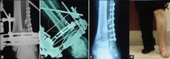
Figure 3.

(a) Postoperative X-ray of ankle joint with leg bones showing lateral translation of the distal fragment (b) The lateral translation was corrected by means of a Olive-wire in same patient (c) X-ray anteroposterior view of ankle joint and clinical photograph (d) showing fracture union and fixator has been removed; patient weight bearing (at 5 months followup)
