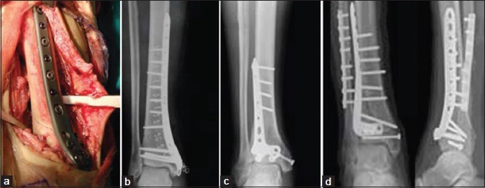
Figure 4.

(a) Peroperative photograph showing anteromedial approach to the distal tibia used to perform open reduction and internal fixation with a medial plate (b) Postoperative X-ray leg bones with ankle joint showing the reduction achieved. (c and d) X-rays of leg bones with ankle showing A.L.P.S. Anterolateral plates, without fibular plate (c) and with fibular plate (d)
