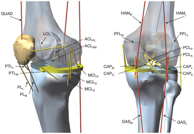
Figure 2.
Schematic of the knee model showing muscles and ligaments: quadriceps, medial and lateral hamstrings, and medial and lateral gastrocnemius muscles (QUAD, HAMM, HAML, GASM, and GASL, respectively); the anteromedial and posterolateral bundles of the anterior cruciate ligament (ACLAM and ACLPL, respectively); the anterior and posterior bundles of the posterior cruciate ligament (PCLA and PCLP, respectively); the lateral collateral ligament and the superficial, deep, and posterior medial collateral ligament (LCL, MCLS, MCLD, and MCLP, respectively); the medial and lateral patellofemoral ligament and the medial and lateral patellotibial ligament for the anterior knee capsule (PFLM, PFLL, PTLM, and PTLL, respectively); the medial, lateral, oblique popliteal, and arcuate popliteal fiber for the posterior knee capsule (CAPM, CAPL, CAPO, and CAPA, respectively); and the medial and lateral patellar ligament (PLM and PLL, respectively).

