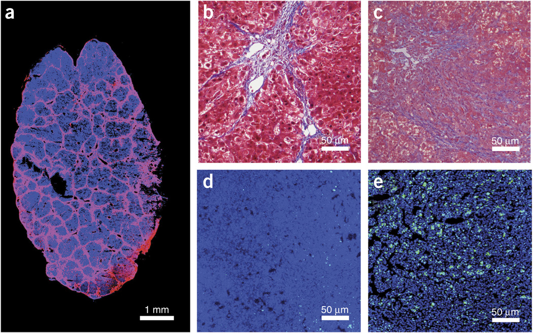
Figure 2.
Microscopic appearance of hepatocellular carcinoma in mice with cirrhotic liver. (a) Immunofluorescence staining of collagen I in mice with liver cirrhosis (Laennec 4 score) after 12 weeks of CCl4 treatment. In blue, DAPI cell nuclear counterstaining. (b,c) Masson’s trichrome staining of liver tissue from C3H (b) or Stk-mutant (c) mice with cirrhosis. (d,e) Representative images of CD8+ T lymphocyte infiltration (green, anti-CD8 antibody) in HCA-1 (d) and Stk-mutant tumors (e) with DAPI (VECTASHIELD) for nuclear counterstaining, in blue. Institutional regulatory board permission from MGH IACUC was obtained for all procedures performed within this protocol.

