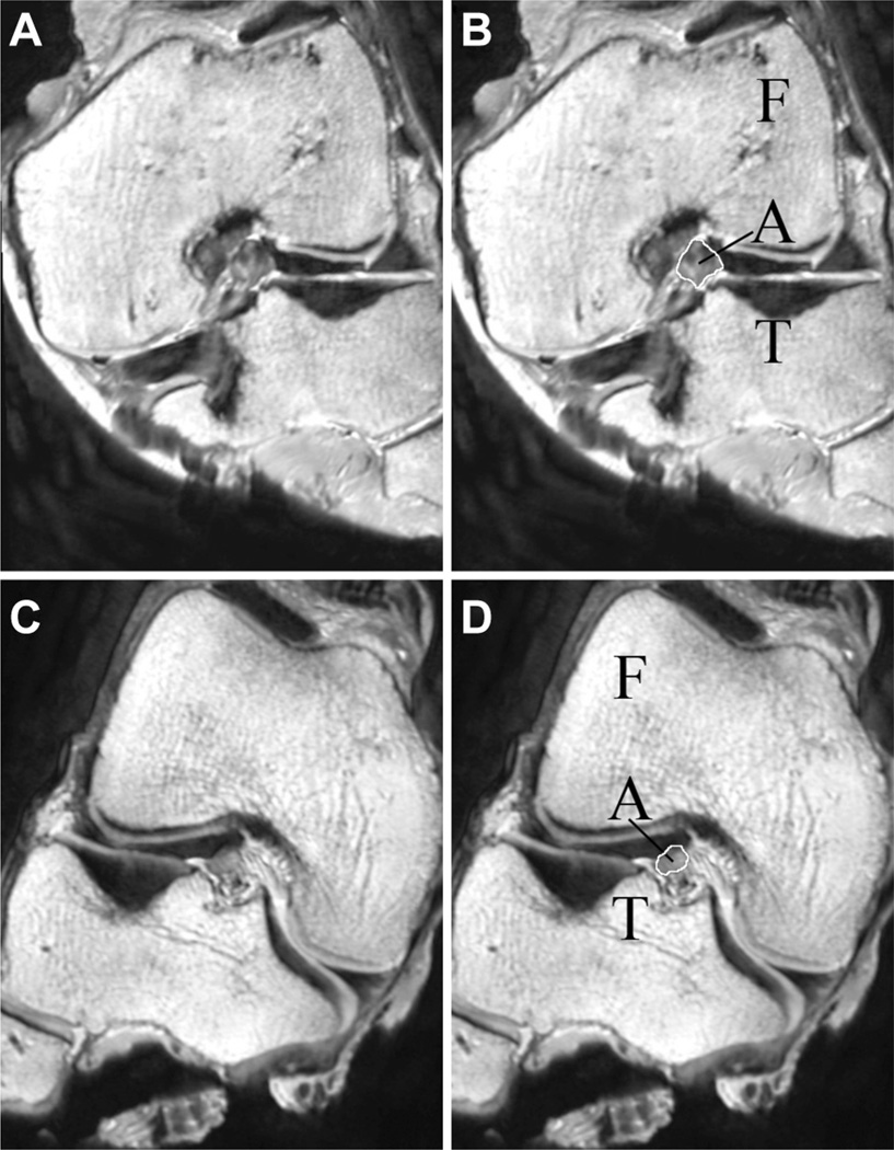
Figure 3.
(A) Oblique-axial view of the left knee of male specimen #33400 at 30% of the ligament’s length from the tibial insertion. (B) The femur (F), tibia (T), and anterior cruciate ligament (ACL, A) are labeled on male specimen #33400. The cross-sectional area of the ACL is outlined in white. (C) Oblique-axial view of the right knee of female specimen #33272 at 30% of the ligament’s length from the tibial insertion. (D) The femur (F), tibia (T), and ACL (A) are labeled on female specimen #33272. The cross-sectional area of the ACL is outlined in white.

