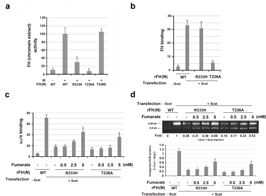
Figure 5. Fumarate produced by chromatin-associated FH promotes NHEJ.
The data represent the mean ± SD (n=3 independent experiments). c, d, * stands for P < 0.05 between the indicated samples and the WT counterparts without adding exogenous monoethyl-fumarate.
a, Chromatin extracts of thymidine double block-synchronized U2OS cells were collected 1 h after IR. Relative FH activity was measured. a, * stands for P < 0.05 between the indicated samples and the WT counterpart.
b, DR-GFP-expressing U2OS cells with depleted FH and reconstituted expression of the indicated FH proteins were harvested 30 h after transfection with a vector expressing I-SceI. ChIP analyses with an anti-FH antibody were performed.
c, DR-GFP-expressing U2OS cells with depleted FH and reconstituted expression of the indicated FH proteins were incubated with the indicated concentration of monoethyl-fumarate for 20 h after I-SceI transfection. ChIP analyses with an anti-Ku70 antibody were performed.
d, DR-GFP-expressed U2OS cells with depleted FH and reconstituted expression of the indicated FH proteins were incubated with the indicated concentration of monoethyl-fumarate for 20 h after I-SceI transfection. An NHEJ analysis was performed. A representative image of the PCR products digested by I-SceI and BcgI is presented.

