Figure. 1.
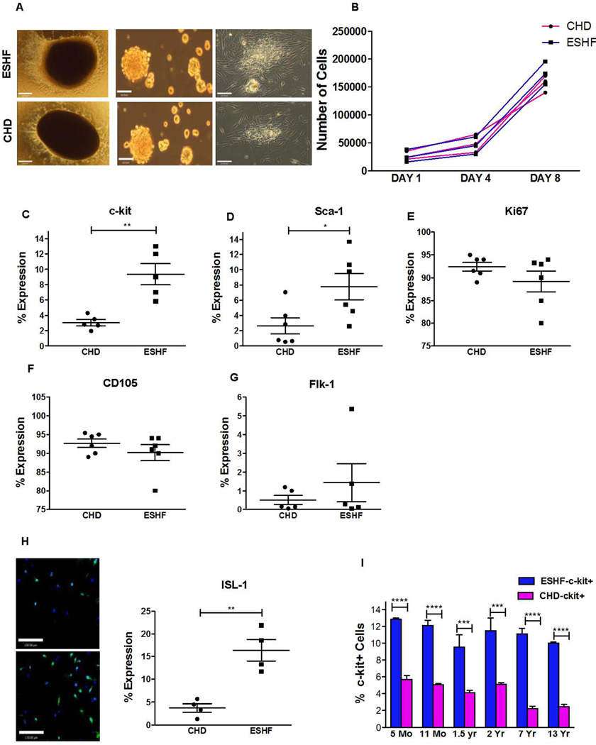
Characterization of ESHF and CHD derived CDCs. (A,i–vi) Generation of cardiospheres from RAA explant cultures. (i & iv) Adherent tissue explant present at one week. (ii & v) Phase-bright cells were removed and replated them in cardiosphere-growing medium–induced cardiosphere formation (iii & vi). Cardiospheres expanded to a monolayer of hCDCs (Scale bar = 120µm). (B) Population doubling over time between ESHF and CHD derived hCDCs was similar. (C–G) Antigenic phenotype of ESHF and CHD derived CDCs demonstrated higher expression of c-kit+ (**P=0.0079) and Sca-1 (*P=0.041) and not Ki67, CD105, Flk-1. (H) Elevated ISL-1 expression was seen in ESHF derived CDCs when compared to CHD derived CDCs determined by immunofluorescence (**P<0.01) (scale bar = 120µm). (I) A higher number of c-kit+ cells were present in ESHF derived CDCs than in CHD derived CDCs (***P<0.0001, ****P<0.0001). Data are analyzed by non-parametric t-test (Mann-Whitney’s analysis) and presented as mean ± SEM.

