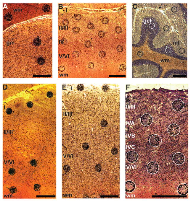
Figure 1.
Nissl-stained sections of tissue with sinapinic acid matrix spots. A: caudate nucleus; Chimpanzee 2. B: somatosensory cortex; Chimpanzee 3. C: cerebellum; Human 7. D: anterior cingulate cortex; Human 1. E: primary motor cortex; Human 7. F: primary visual cortex; Chimpanzee 2. The sections are 10 μm-thick and mounted on a metal plate. Because Nissl stain is applied after the application of the matrix and mass spectrometry (MS) is performed, some matrix crystals move from their original locations. Circles are superimposed on the original positions of the matrix spots on the sections from V1 and CB. Neocortical layers and white matter (wm) are labeled in ACC, M1, S1, and V1. The gray matter (gm) of the CN is labeled as well as the surrounding wm. The granule cell layer (gcl), molecular layer (ml), and wm are identified in the CB. Each scale bar is 500 μm in length. The brightness and contrast of the panels were adjusted.

