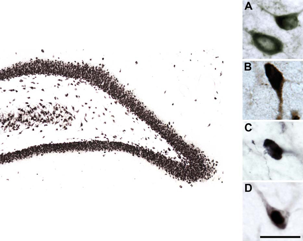
Figure 1.
High-magnification photomicrographs of GAD67-, SOM-, NPY-, and NeuN-immunoreactive hilar neurons. Representative GAD67 (A)-, SOM (B)-, NPY (C)-, and NeuN (D)-immunoreactive hilar neurons viewed at high magnification. Larger image depicts NeuN-immunoreactive neurons in the dentate gyrus. Scale bar = 25 µm in D (applies to A–D); 200 µm for larger image.

