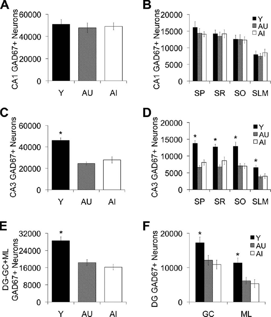
Figure 4.
GAD67-positive interneuron counts in hippocampal subfields CA1, CA3, and DG. A,B: Number of CA1 GAD67-positive neurons remains stable across the adult rat life span regardless of cognitive status. C,D: GAD67-positive neurons are reduced in AU and AI rats in the CA3 hippocampal subregion. E,F: GAD67-positive neurons are reduced in AU and AI rats in the DG moleculare (ML) and granule cell (GC) layers. Numbers of GAD67-positive neurons in the dentate hilus are depicted in Figure 5A,B. Error bars represent ± SEM. Y, young; AU, aged unimpaired; AI, aged impaired; GAD67, glutamic acid decarboxylase-67; SP, stratum pyramidal; SR, stratum radiatum; SO, stratum oriens; SLM, stratum lacunosum-moleculare. *P < 0.05.

