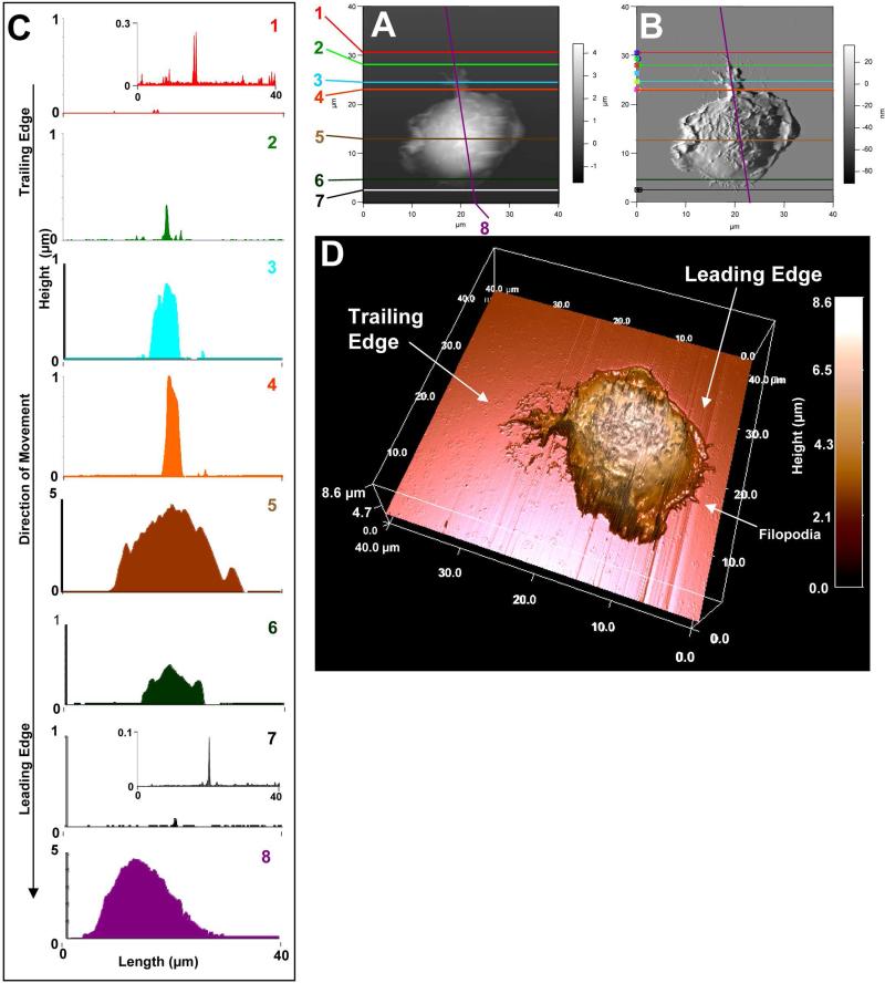
Figure 2.
Membrane features associated with cell movement such as filopodia, and fine structures in the leading and trailing edges. [A] and [B] represent AFM topographies and the corresponding deflection images, respectively. BMMCs were stained with CFSE, sensitized overnight with anti-DNP-IgE and then spun onto a glass coverslip coated with DNP-BSA. After 30 min incubation, cells were fixed with 3.7% formaldehyde for 30 min. [C] is a 3D rendering of the topographic image. Cursor profiles crossing the filopodia shown in [A] and [B] are shown in the left column to provide corresponding height data at a given cursor line. AFM images of the fixed cell was conducted in contact mode with CSC38 cantilevers (k = 0.03 N/m, MikroMasch lever B).

