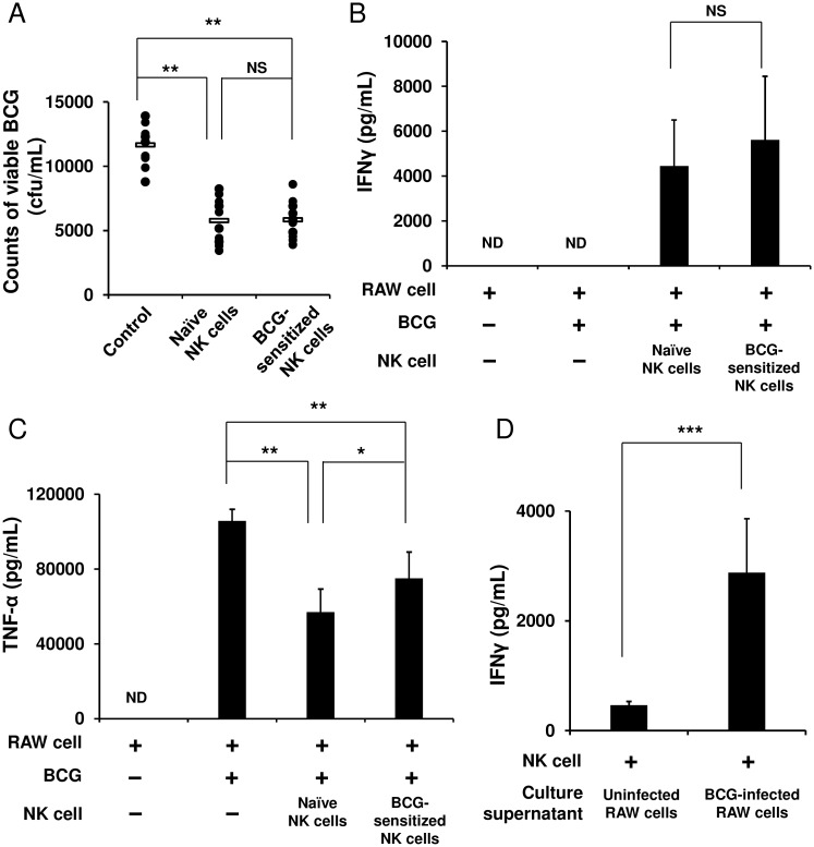
Fig 3. NK cells markedly enhance the ability of macrophages to eradicate BCG.
RAW 264.7 murine macrophage cells were infected with BCG (MOI = 3) at 37°C for 2 h, washed with PBS three times, and then plated at 1 × 10⁶ cells/mL in a 12 well plate. Purified NK cells (3 × 10⁵), which had been stimulated with BCG (MOI = 1) at 37°C for 4 h, were added to the BCG-infected RAW cell culture. As controls, unstimulated naïve NK cells were added to the BCG-infected RAW cell culture, and the BCG-infected RAW cells alone were additionally prepared. Forty eight hours later, these cells were harvested and lysed with 1 mL of 0.067% SDS solution. Serial dilutions were plated on Middlebrook 7H10 agar plates, and 3 weeks later, the number of bacterial colonies grown on the agar plates were counted (A). As in (A), IFNγ (B) and TNF-α (C) in the culture supernatants were measured by ELISA. Purified naïve NK cells were cultured in medium supplemented with either the culture supernatant of the BCG-infected RAW cells or uninfected control RAW cells at a ratio of 1:1 at 37°C for 24 h, and IFNγ in the culture supernatants was measured using ELISA (D). The data are presented as mean ± standard deviation, and p values < 0.05 were considered statistically significant. Similar results were obtained in three independent experiments. Horizontal bar in (A), mean value; *p < 0.05; **p < 0.01; ***p < 0.0001; NS, not significant.

