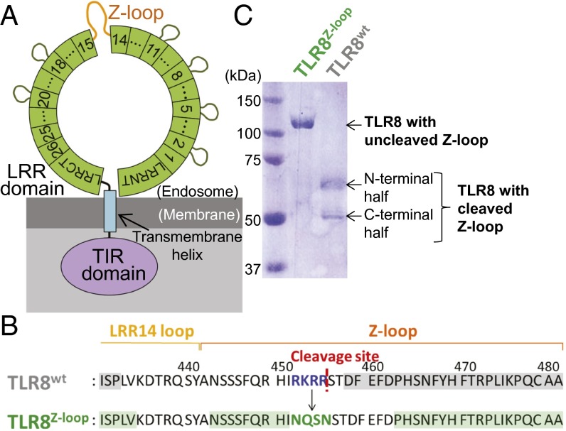
Fig. 1.
Z-loop cleavage of human TLR8. (A) Schematic representation of the domain organization of human TLR8. The ring (green), rectangular box (blue), and oval (purple) represent the extracellular LRR domain, transmembrane helix, and intracellular TIR domain, respectively. LRRs are indicated by numbered boxes. The Z-loops are shown in orange. (B) Sequence of TLR8wt (Top) and TLR8Z-loop (Bottom). The cleavage site in TLR8wt is shown as a red dashed line. Mutation sites are shown in blue (TLR8wt) and green (TLR8Z-loop). The residues visible in the electron density maps are highlighted in gray (TLR8wt) and green (TLR8Z-loop). (C) SDS/PAGE (10% polyacrylamide) analysis of the purified TLR8Z-loop (Right) and TLR8wt (Left) under reducing conditions.

