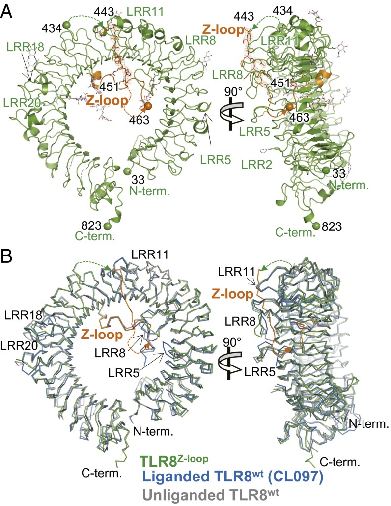
Fig. 3.
Crystal structure of TLR8Z-loop. (A) Front (Left) and side (Right) views of TLR8Z-loop structure. The LRR structure and Z-loop are shown in green and orange, respectively. N-glycans and disulfide bonds are shown as gray and yellow sticks, respectively. The N and C termini of each fragment are shown as spheres. The dashed lines show unmodeled regions in the structure. (B) Superposition of TLR8Z-loop with TLR8wt. TLR8Z-loop (green), one protomer from unliganded TLR8wt dimer (gray; PDB ID code 3W3G), and one protomer from liganded TLR8wt dimer (blue, TLR8/CL097; PDB ID code 3W3J) are superposed and shown in Cɑ trace models. The Z-loop in TLR8Z-loop is shown in orange.

