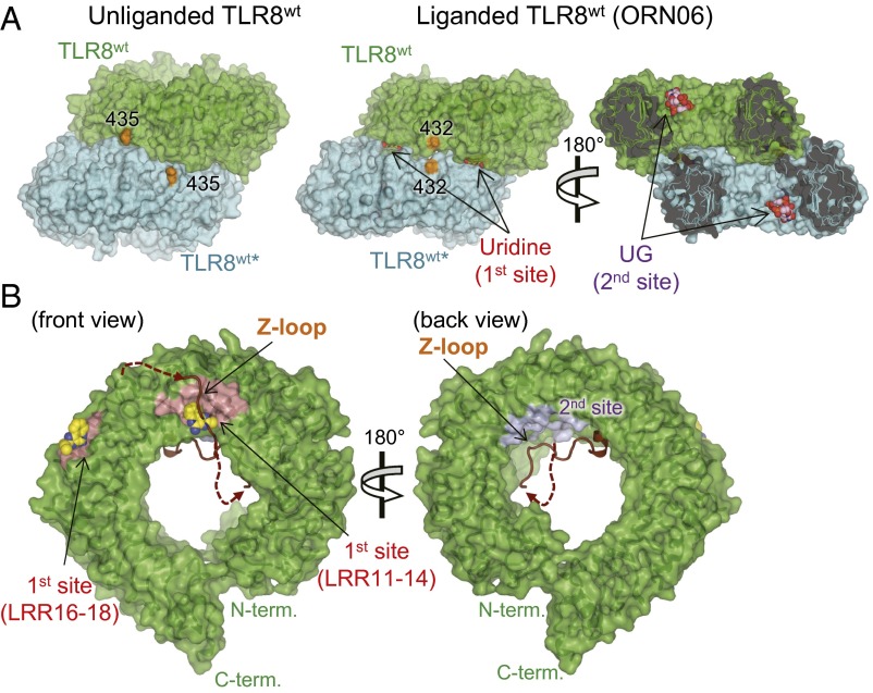
Fig. 5.
Surface representations of TLR8Z-loop and TLR8wt. (A) Surface representations of the unliganded TLR8wt (Left) and liganded TLR8wt (Right, TLR8/ORN06; PDB ID code 4R07). TLR8wt protomers are shown in green (TLR8wt) and cyan (TLR8wt*). Uridine (first site) and UG (second site) molecules are shown in a space-filling representation. The C atoms of the ligands are colored yellow (uridine) or purple (UG), and the O, N, and P atoms of the ligands are colored red, blue, and orange, respectively. The end of the LRR14 loop is in orange. (B) Surface representations of front (Left) and back (Right) views of TLR8Z-loop. The LRR14 loop and the Z-loop are shown in brown in a cartoon model. The first and second sites are pink and purple, respectively. The chemical ligand (CL097) in TLR8WT/CL097 shown as a sphere is overlaid on TLR8Z-loop structure.

