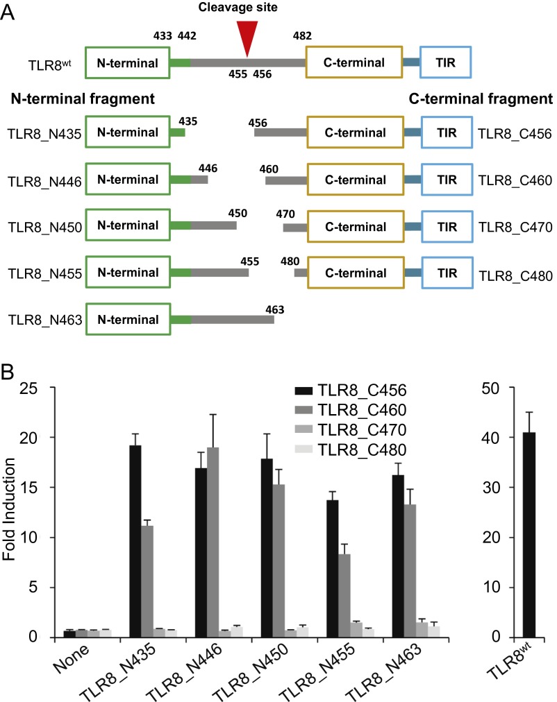
Fig. S1.
Reconstruction of functional TLR8 by N- and C-terminal fragments. (A) Schematic representation of various N- and C-terminal deletion mutants of human TLR8 used in the present study. The numbers denote the amino acid positions in each construct. The LRR14 loop (residues 433–441) and Z-loop (residues 442–481) are depicted in green and gray lines, respectively. (B) NF-κB activation in HEK293T cells expressing indicated human TLR8 fragments and TLR8WT. The response was assessed using an NF-κB–dependent luciferase assay stimulated by 5 mM uridine and 25 μg/mL ssRNA40 complexed with DOTAP. Data represent the mean fold induction of NF-κB activity, calculated as the RLU of stimulated cells divided by the RLU of nonstimulated cells. Data from six independent samples are shown.

