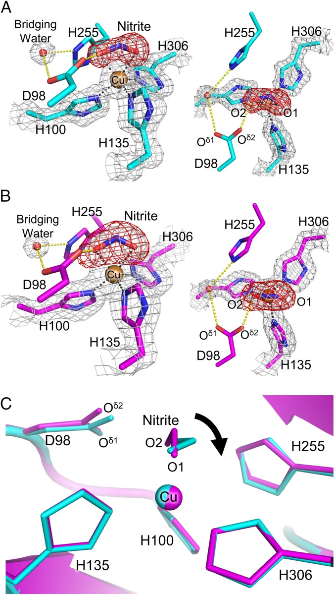
Fig. 1.
NO2− binding in NC structures. (A) T2Cu site in the SRX NC structure (molecule A). The sigma-A–weighted 2Fo–Fc (1.5 σ) and omit Fo–Fc (6.5 σ) maps are shown as gray and red meshes, respectively. H-bonds (yellow) and coordination bonds (black) are represented by dashed lines. C, N, O, and Cu atoms are colored cyan, blue, red, and brown, respectively. (B) T2Cu site in the SFX NC structure (molecule A). The sigma-A–weighted 2Fo–Fc (1.0 σ) and omit Fo–Fc (4.5 σ) maps are shown as gray and red meshes, respectively. H-bonds and coordination bonds are represented as in A. C, N, O, and Cu atoms are colored magenta, blue, red and brown, respectively. (C) Comparison between the SFX NC (magenta) and SRX NC (cyan) structures.

