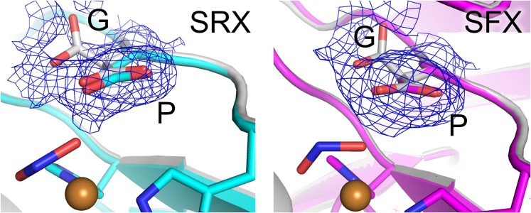
Fig. 5.
Conformations of Asp98. The sigma-A–weighted 2Fo–Fc maps (0.2 σ) are shown as blue meshes. The SRX NC and SFX NC structure are shown in cyan and magenta, respectively. The structure showing G and P conformations of Asp98 (PDB ID code 2BWI) (21) is colored white.

