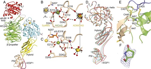
Fig. 1.
LFA-1 headpiece structure. (A) Representative LFA-1 headpiece structure [crystal in Mg formate, Protein Data Bank (PDB) ID code 5E6U]. Ribbon cartoon with disulfides as gold sticks, glycans as white sticks with red oxygens, and Ca2+ and Mg2+ as gold and silver spheres, respectively. βI domain metal binding sites of (B) LFA-1 crystal in Mg formate and (C) internally liganded αXβ2 crystal (PDB ID code 4NEH). (D) β-subunit domain orientation differences shown after superposition on the βI domain of the two most dissimilar LFA-1 headpiece examples (cyan, PDB ID codes 5E6S and 5E6U), rerefined closed–bent αXβ2 ectodomain (orange, PDB ID code 5ES4), and internally liganded bent αXβ2 ectodomain (red). (E) Interface between the PSI (green) and I-EGF1 (wheat) domains in PDB ID code 5E6U. (F) N-terminal pyroglutamic acid (PDB ID code 5E6V). Mesh shows 1σ 2Fo–Fc density.

