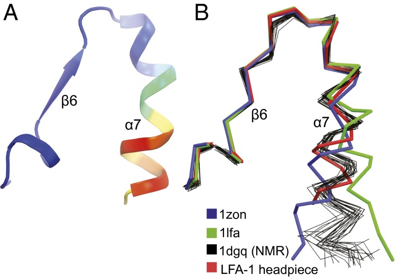
Fig. 4.
αI domain α7-helix flexibility. Structures are superimposed on the αI domain with only the C-terminal portion of αI domain shown. A and B are in identical orientations. (A) Ribbon cartoon of the 2.15 Å αLβ2 structure colored by the Cα atom B factor from low (blue) to high (red). (B) Comparisons of isolated αL αI domain crystal structures (PDB ID codes 1LFA and 1ZON) (6, 7), thick ribbon traces; isolated αI domain NMR structure (PDB ID code 1DGQ) (11), thin ensemble ribbon traces; and αL αI domain in headpiece crystal structure, thick ribbon trace.

