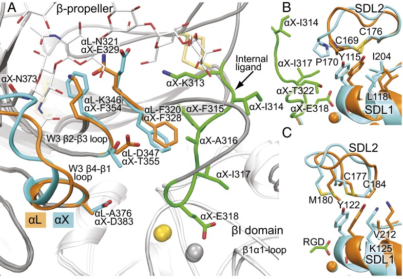
Fig. 5.
The internal ligand-binding pocket and concerted movement of SDL1 and SDL2. (A) The internal ligand-binding pockets of the αLβ2 headpiece and internally liganded, cocked αXβ2 ectodomain after superimposition on the β-propeller and βI domains. The conserved Phe and pocket residues and backbone that differ between αL and αX are shown with orange (αL) or cyan (αX) side chains and ribbon cartoon. The internal ligand of αX is similarly shown in green. Otherwise, α and β subunits are shown in gray and white ribbon cartoon, respectively. Metal ions are spheres in gold (SyMBS Ca2+) and silver (MIDAS Mg2+). (B and C) SDL1-enforced movement of SDL2 in β2 integrins (B) and αIIbβ3 (C). Closed αLβ2 (PDB ID code 5E6S) and αIIbβ3 (PDB ID code 3T3P) conformations are in orange, whereas internally liganded, cocked αXβ2 (PDB ID code 4NEH) and open αIIbβ3 conformations (PDB ID code 2VDR) are in cyan (16, 30). Structures are in identical orientations and show ribbon cartoon and key side chains in stick.

