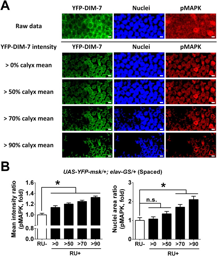
Fig. S7.
Nuclear accumulation of activated MAPK in MB nuclei by overexpression of YFP-fused DIM-7 at hour 8 after spaced training. (A) Representative images of a part of MB nuclei area in flies with acute overexpression of YFP-fused DIM-7 at hour 8 after spaced training. YFP–DIM-7 signals are shown as green, nuclei signals as blue, and pMAPK signals as red. Signals of a part of MB nuclei area are shown as raw data (above the black line); signals of the same part are shown only within nuclei (below the black line), which are masked by different YFP–DIM-7 intensity (changed with intensity above 0%, 50%, 70%, or 90% mean intensity of YFP–DIM-7 in calyx). (B) Statistical analysis as reflected in mean intensity ratio (averaged intensity of nuclear pMAPK staining, Left) and nuclei area ratio of pMAPK (number of nuclei with strong pMAPK signal, Right). Considering uneven overexpression within the population of MB neurons, different subgroups of nuclei with strong overexpression of YFP-fused DIM-7 (above 0%, 50%, 70%, or 90% of calyx mean intensity) are included. Acute expression was induced by RU486 feeding (RU+). No RU486 feeding served as control. Bars, mean ± SEM (n = 7–12); *P < 0.05. n.s., no significance.

