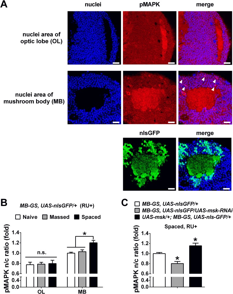
Fig. S8.
DIM-7 affects nuclear accumulation of pMAPK in MB neurons bidirectionally at hour 8 after spaced training. (A) Representative images of pMAPK and nuclei signals in optic lobe (Top) and MB (Middle and Bottom). Images include nuclear staining (Left), pMAPK staining or nlsGFP signal (Middle), and merged staining (Right). Apparently strong immunostaining of pMAPK was observed in MB nuclei (white arrowhead), but not in optic lobe. (Scale bars, 10 μm.) (B) Statistical analysis as reflected in mean intensity ratio (averaged intensity of nuclear pMAPK staining) in optic lobe (OL) and MB in flies with naive state or at 8 h after massed and spaced training. The ratios of all groups were normalized to MB group with naive condition. (C) Statistical analysis as reflected in mean intensity ratio (averaged intensity of nuclear pMAPK staining) in flies with acute knockdown (shaded bar) or overexpression of DIM-7 (solid bar) in contrast to control group (open bar) at 8 h after spaced training. Acute expression was induced by RU486 feeding (RU+). Bars, mean ± SEM (n = 6). *P < 0.05. n.s., no significance.

