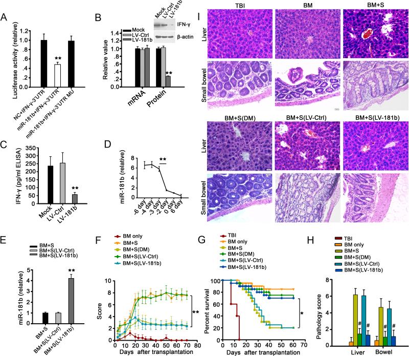
FIGURE 3. MiR-181b alleviates aGVHD by targeting IFN-γ in mice.
(A) Luciferase activity of reporter bearing wide type or mutant (MU) 3'UTR of IFN-γ co-transfected into HEK293T cells with murine miR-181b. (B) Relative levels of IFN-γ mRNA and protein in activated CD4+ T cells isolated from spleens of mice infected with LV-181b or LV-Ctrl; CD4+ T cells were activated by PMA and ionomycin. The mRNA and protein levels in mock-infected cells as negative control were set to 1. ** p < 0.01. (C) Expression of IFN-γ protein in mice was quantified by ELISA. ** p < 0.01. (D) Relative expression levels of miR-181b in plasma of recipient mice with aGVHD on days around the onset of aGVHD. Day 0 represented the day of aGVHD onset. The level of miR-181b was significantly decreased prior to development of aGVHD. ** p < 0.01. (E) Expression level of miR-181b in plasma of mice receiving T lymphocyte-depleted bone marrow cells and splenocytes (BM+S), LV-Ctrl (BM+S (LV-Ctrl)) or LV-181b infected splenocytes (BM+S (LV-181b)). ** p < 0.01. (F) Clinical scores of different groups of recipient mice after transplantation. ** p < 0.01. (G) Survival rate of different groups of mice after transplantation. * p < 0.05. Scores (H) and H&E stained sections (I) of the liver and bowel samples from different groups of mice at day 12 after transplantation. ** p < 0.01, # p < 0.001, compared to group BM+S. All values (A-H) represent mean±SD. Data are from three independent experiments, representing 6 mice per group (B-H).

