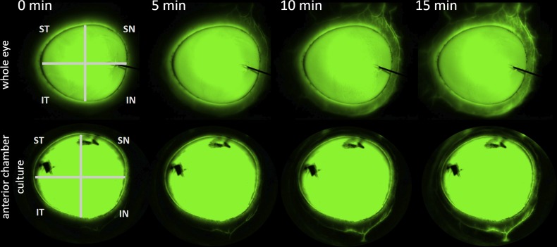
Fig 3. Example canalogram time lapse of a whole eye (top) and an anterior chamber culture (bottom).
Time is indicated in minutes at the top. IN = inferonasal, SN = superonasal, ST = superotemporal, IT = inferotemporal quadrant. Dye marks which were used during pilot experiments can be seen on the cornea blocking fluorescence.

